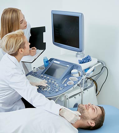
An Echocardiogram or “Echo,” otherwise known as an ultrasound of the heart, uses high-frequency sound waves to image the heart. The exam is performed by an accredited cardiac sonographer and takes on average 30-45 minutes, depending on an individual’s image quality and the complexity of the findings. In a transthoracic echocardiogram, images are obtained by applying cool gel to a transducer which is then moved over the patient’s chest wall, in between and around the ribs, to visualise the heart. Firm pressure may need to be applied to the chest wall in order to see the heart but the sonographer will do their best to minimise any discomfort. During the exam, you will be asked to lie in different positions and may need to hold your breath at certain times. No preparation is required for this ultrasound. An echocardiogram provides very detailed information on the size of the heart chambers and great vessels (aorta and pulmonary artery), the function of the heart muscle and heart valves, as well as any fluid around the heart.

The sonographer will report their findings and within the week, images will be reviewed by one of our cardiologists who then finalise the echo report. Findings are of great use to the treating cardiologist in diagnosing heart conditions and managing symptoms; or your general practitioner / referring physician may refer you to a cardiologist, based on your echo results. Alongside the ECG (electrocardiogram), an echo is the most common cardiac diagnostic test used to assess the heart.